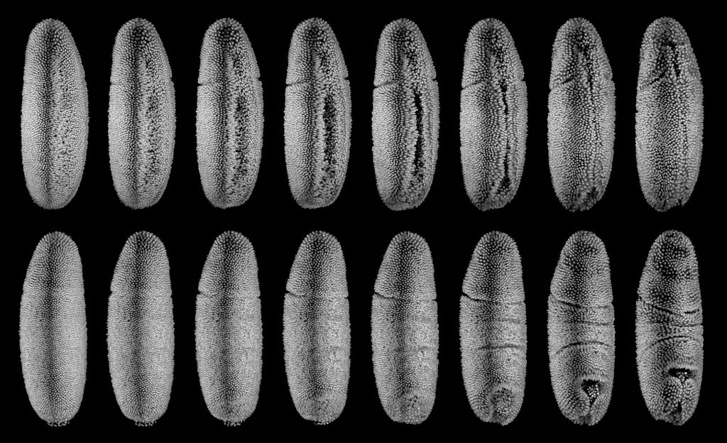
Drosophila melanogaster gastrulation

Perspective view: Ventral side
3D stereoscopic view: Ventral side
3D anaglyphic view: Ventral side
Perspective view: Dorsal side
3D stereoscopic view: Dorsal side
3D anaglyphic view: Dorsal side
Perspective view: Ventral side
3D stereoscopic view: Ventral side
3D anaglyphic view: Ventral side
Perspective view: Dorsal side
3D stereoscopic view: Dorsal side
3D anaglyphic view: Dorsal side
Snapshots from a live recording of Drosophila melanogaster embryonic development using a 3D light-sheet microscope. Visible structures are labeled cell nuclei. Data was recorded, post-processed and visualized by Stefan Günther at EMBL.