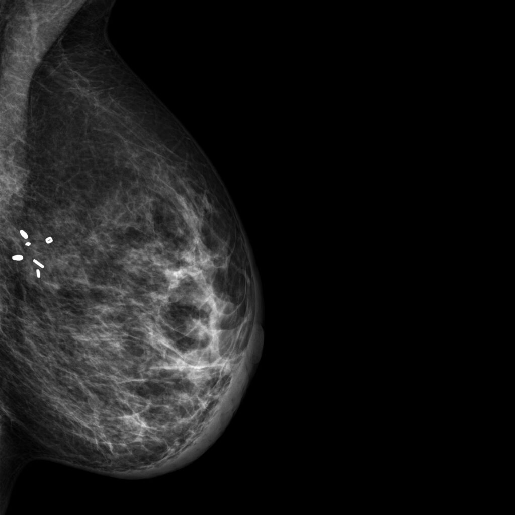
Mammogram with implanted surgical marker
A typical mammogram is shown. X-rays are absorbed and scattered in calcium rich tissue which appears white. Mammograms are an overlay of all structures along the radiation beam, thus lacking depth information. The bright tissue on the left is the pectoralis muscle. Implanted surgical markers are visible and extremly bright in mammograms. At the breast edge at the right the nipple is visible. Another landmark for a perfect mammogram is the visible and non-obscured inframammary fold (IMF) at the lower left.
Courtesy of Wikimedia Commons.
CC-BY-SA-4.0, https://commons.wikimedia.org/wiki/File:Mammogram_shows_BioZorb_surgical_marker_implanted_in_the_breast.jpg